Magnetic tensiometer
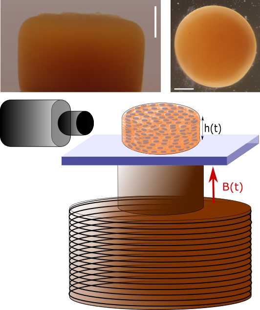
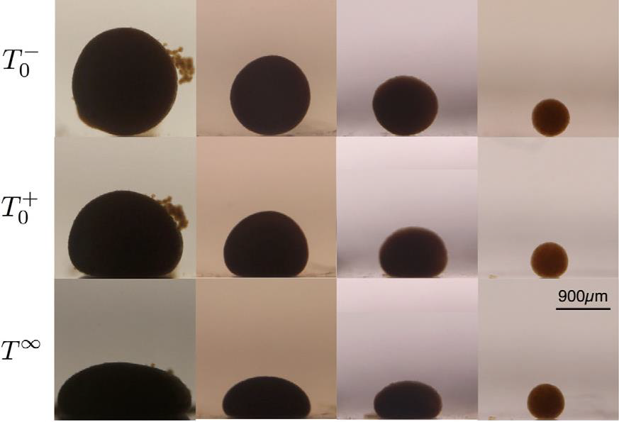
Magnetically responsive cells can be remotely stimulated using external magnetic fields, acting as mechanical actuators within active tissues. By applying controlled magnetic forces, we can generate tissue-scale deformations without direct mechanical contact. The approach of a magnet produces a body force on the magnetic cells, analogous to an effective supergravity, and thereby deforms magnetic multicellular aggregates.
This principle forms the basis of our magnetic tensiometer, an approach that combines active matter concepts with the physics of soft tissues. By analyzing the deformation of multicellular aggregates under magnetic forces, we use a fluid-mechanics framework to characterize their mechanical properties. In particular, the flattened profile adopted by the aggregates provides access to an effective surface tension, allowing us to quantify tissue cohesion and mechanical behavior.
The magnetic tensiometer therefore provides a non-invasive and remotely controlled method to probe the mechanics of living tissues, while simultaneously manipulating the forces acting within them. It offers a unique tool to investigate how cellular activity and tissue mechanics contribute to the organization and emergent properties of active tissues.[1].
Tissue surface tension is governed by both cell–cell adhesion and the intrinsic mechanical properties of cells. Changes in the cytoskeleton can therefore strongly influence the mechanical state of tissues. In particular, our work has shown that remodeling of actin and intermediate filament networks can modify tissue surface tension, highlighting the role of the cytoskeleton in regulating the mechanical cohesion of multicellular tissues [2].
Importantly, tissue surface tension is also altered during malignant transformation and epithelial–mesenchymal transition (EMT). These changes in tissue cohesion and mechanics are associated with alterations in cell adhesion, migration, and invasive behavior. Measuring tissue surface tension therefore provides a quantitative approach to characterize changes in the mechanical state of tissues during tumour progression and suggests its potential as a biophysical marker of tumour state and invasiveness [3].
Magnetic rheometer
By applying magnetic forces with controlled timing and frequency, we can probe the dynamic mechanical properties of multicellular aggregates. This approach allows us to investigate how model tissues deform, relax, and remodel in response to mechanical stimulation, providing insights into their viscoelastic and active behavior.
Interestingly, although multicellular aggregates display mechanical responses reminiscent of those observed at the single-cell scale, they are surprisingly much softer than individual cells [4]. This unexpected collective behavior highlights how cell–cell interactions and cellular organization give rise to emergent tissue mechanics.
Our magnetic rheometer provides a unique tool to explore the dynamics of active tissues, from cellular force generation to tissue-scale remodeling. We are particularly interested in how these mechanical properties evolve during tumour progression and invasion. Changes in cell adhesion, cytoskeletal organization, and active force generation can profoundly modify tissue cohesion and deformability, enabling cell rearrangements and invasive behavior. By combining controlled mechanical stimulation with model tumour tissues, we aim to understand how tissue dynamics and active mechanical processes contribute to the transition from a cohesive tumour to an invasive state.
[1] Mazuel F. et al. (2015) Phys. Rev. Lett.
[2] Nagle et al. (2022) Elife
[3] Nagle I. et al. (2022) Frontiers in Cell and Dev. Biol.
[4] Mary G. et al. (2022) Phys. Rev. E